UNEC Journal of Engineering and Applied Sciences Volume 4, No 1, pages 83-91 (2024) Cite this article, ![]() 2072 https://doi.org/10.61640/ujeas.2024.0510
2072 https://doi.org/10.61640/ujeas.2024.0510
The genus Curtobacterium is a member of the Mcrobacteriaceae family and the phylum Actinomycetota (formerly known as Actinobacteria) [1]. Certain Curtobacterium strains are plant pathogenic [2], while many are categorized as either rhizobacteria that promote plant development or endophytes that potentially reduce the host’s abiotic stresses [3-5]. The C. flaccumfaciens species includes a number of closely associated plant pathogens of economic and agricultural significance that are divided into different pathovars with varying host levels. These pathovars include C. flaccumfaciens pv. betae (Betae vulgaris), C. flaccumfaciens pv. flaccumfaciens (Phaseolus vulgaris), C. flaccumfaciens pv. basellae (Basella alba) and many others [6-7]. C. flaccumfaciens is gram-positive, with an irregular form of short rods [2], [8]. The stains are orange and yellow in color, motile and never generate endospores, aerobic, oxidase-negative, catalase-positive [9-10]. In fact, the majority of research on Curtobacterium emphasizes how economically significant a plant pathogen it is [11,12]. According to reports on five continents, the most well-studied pathovar, C. flaccumfaciens pv. flaccumfaciens, is the cause of bacterial wilt in dry beans [13,14]. Even among an individual host, the disease exhibits a high level of phenotypic and genetic variability [15,16]. The 30S subunit of a bacterial ribosome, also known as SSU rRNA, is made up of 16S ribosomal RNA, commonly referred to as 16S rRNA. It provides the majority of the SSU structure and binds to the Shine-Dalgarno sequence [17]. Since the 16S rRNA gene is highly conserved throughout several species of bacteria, it is employed for phylogenetic analyses. Due to the discovery that 16S rRNA sequences from distantly associated bacterial lineages have identical functions, it is proposed that the 16S rRNA gene can serve as a credible molecular clock [18,19]. The 16S rRNA gene sequences feature hypervariable sites alongside highly conserved regions that may deliver species-specific hallmark sequences helpful for identifying bacteria [20]. As a quick and affordable solution for phenotypic approaches to bacterial identification, the sequencing of 16S rRNA genes has consequently gained popularity in microbiology studies [21]. One of the oldest cultivated crops, Olea europaea, needs less in regards to the use of agricultural supplies [22]. Given that olive-associated bacteria have demonstrated various plant growth promoting features both in vitro [23] and in vivo [24], it is likely that interaction with desirable soil microorganisms also contributes to the adaptation of olives. Due to the lack of studies that show the relationship between C. flaccumfaciens and the olive (O. europaea) as a host, the current manuscript aimed to investigate the phenotypic, molecular, and biochemical properties of this bacterium.
Samples collection
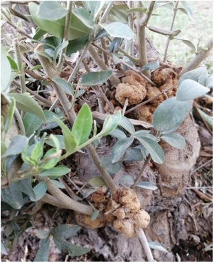
Approximately 100 samples were collected from the nodes of O. europaea trees (figure 1) from different regions of Nineveh Governorate, including University of Mosul, Al-Arabi district, Al-Fadhiliya, and Bashiqa using sterile bottles during the period 1/6-8/2023. Later, the samples were labelled and transferred to the laboratory, and they were washed with tap water to remove dust and contaminants. After that, the samples were sterilized with 10% sodium hypochlorite for 10 min [25], then the nodes were washed well with distilled water D.W to remove the bleach substance.
Sterilization of culture media and solutions
The synthetic and ready-made media and solutions that are not affected by heat are sterilized using an autoclave at a temperature of 121 °C under pressure 2 bar for 20 min [26]. Tools and surfaces were sterilized with 70% alcohol.
Isolation and cultivation of C. flaccumfaciens
The nodes were crushed thoroughly with a ceramic mortar, and immersed in physiological normal saline to maintain bacterial viability. The bacteria were incubated overnight at 28 °C for 24 h. Nutrient agar medium was prepared according to the instructions of the manufacturer of Neogen/ UK (Spanish). This was done by dissolving 28 g of nutrient agar in 1 liter of D.W. The overnight cultures that were grown on petri dishes containing NA medium were incubated at 28 °C for 72 h, and examined for growth and color change [2].
Morphological analysis
The phenotypic characteristics of grown colonies on nutrient agar medium were identified, including the shape, size, color and edge of these cells.
Biochemical analysis
Assessment of Gram stain
A swab of bacterial isolates was placed on a clean glass slide, a drop of crystal violet dye was added for 1 min, the slide was washed with dH2O. The iodine was applied to the slide for 1 min, and washed again with dH2O. Then, alcohol was added to the slide for 10 sec, and washed with distilled water. The safranin was added for 1 min. The cells were screened under the light microscope using an oil lens (100x). If the bacteria maintained the crystal violet stain, then the test is positive for gram stain [27].
Oxidase enzyme activity
This reagent was prepared by dissolving 1 g of tetramethyl-p-phenylenediamine dihydrochloride in 100 mL of distilled sterile water [28]. A colony grown on NA medium was transferred onto filter paper using sterile wooden sticks, then a drop of the oxidase reagent was added to it. The color changing to purple within 30 to 60 sec is evidence of enzyme production [29].
Catalase enzyme activity
The 3% hydrogen peroxide solution was prepared from 30% of the original solution [30]. The bacteria cells were placed on a sterile glass slide using sterile wooden sticks, a drop of catalase reagent was added to the slide. The positive test gives air bubbles [28].
Assessment of Motility
A semi-solid medium of 0.5% agar and 8% gelatin was prepared and inoculated with a colony of bacteria using a stabbing method, and incubated at 28 °C for 24 h. The test is considered positive, if the bacterial growth spreads outside the stab line of inoculation [31].
Assessment of antibiotic sensitivity
In order to conduct the antibiotic sensitivity test for C. flaccumfaciens strains using the agar disc diffusion method [32], the following antibiotics were chosen to perform this test: Amikacin, Ciprofloxacin, Trimethoprim, Meropenem, Chloramphenicol, and Gentamicin all with (10 μg), Nitrofurantoin (100 μg), Cefixime (5 μg), and Cefotaxime (30 μg) from Bioanalyse company/ Turkey. The antibiotic discs were placed on petri dishes containing C. flaccumfaciens grown on NA medium. The inhibition zone diameter was measured in millimeters using a caliper.
Molecular analysis
The C. flaccumfaciens genomic DNA was extracted using a bacteria DNA isolation kit (GEE150, GEE1.5K) provided by Geneaid /Taiwan. DNA concentrations were determined using the Nanodrop 2000 [33]. PCR was detected in a 20 μl GoTaq® G2 green master mix provided by Promega corporation/USA. The 27F 5' AGAGTTTGATCMTGGCTCAG 3' and 1522R 5' AAGGAGGTGATCCARCCGCA 3' primers were utilized to amplify the 16S rRNA region [34]. The PCR condition was set up as following: initial denaturation at 95 °C for three minutes, 30 cycles of denaturation at 95 °C for thirty seconds, annealing at 55 °C for thirty seconds, extension at 72 °C for thirty seconds, final extension at 72 °C for seven minutes. The PCR product was run on a 1% agarose (w/v) gel. One hundred bp DNA ladder (New England Biolabs, UK) was utilized as a marker. The PCR products were sent for sequencing to Psomagen company/ USA. The sequences were inspected for homology with deposited sequences in GenBank https://blast.ncbi.nlm.nih.gov/Blast.cgi
Morphological identification
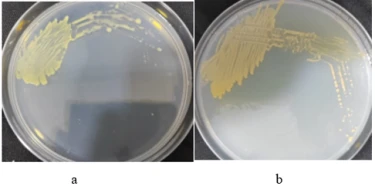
The 5 grown strains on NA medium of C. flaccumfaciens were diagnosed phenotypically in terms of the color, size, shape, and edge. The colonies appeared yellow to orange in color, smooth and shiny with a sharp edge (figure 2).

Figure 2. Phenotypic features of C. flaccumfaciens. a) colonies with yellow color, b) colonies with orange color
Biochemical identification
Gram stain test
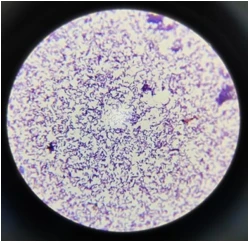
The test was accomplished as previously described above. The bacterial cells were stained in purple or blue color with a short rod shape , this indicates that the cells are gram positive (figure 3).

Figure 3. Microscopic screening of gram staining test of C. flaccumfaciens. The colonies are colored purple or blue, and they are gram positive bacteria
Oxidase enzyme test
A drop of the oxidase reagent was added to a filter paper containing an overnight culture of C. flaccumfaciens. The color does not change to purple, and this is evidence of the absence of an oxidase enzyme (figure 4).
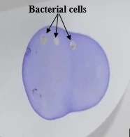
Figure 4. Oxidase test of C. flaccumfaciens . An Oxidase-negative, as no color changes
Catalase enzyme test
A drop of catalase reagent was added to a glass slide containing a colony of C. flaccumfaciens. The appearance of air bubbles on a slide is evidence of the ability of these bacteria to produce catalase enzymes (figure 5).
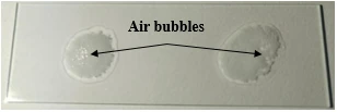
Figure 5. Catalase test of C. flaccumfaciens. A Catalase-positive, as air bubbles appeared on the slide
Motility test
A semi-solid medium was inoculated with bacterial colonies, and kept overnight at 28 °C. The result showed growth spread outside the inoculation stab line (figure 6).
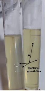
Figure 6. Motility test. A semi-solid medium shows a positive result, as the bacterial growth was spread beyond the stab line of inoculation
Antibiotics sensitivity test
The Amikacin, Ciprofloxacin, Trimethoprim, Meropenem, Chloramphenicol, Gentamicin, Nitrofurantoin, Cefixime, and Cefotaxime antibiotics were used to accomplish the sensitivity test of C. flaccumfaciens. using agar disc diffusion method . The growth inhibition zones in SHGH1, SHGH2, SHGH3, SHGH4, and SHGH5 strains were (2.0, 2.1, 3.1, 2.4, 1.5 mm), (2.8, 2.6, 3.4, 2.8, 0.9 mm), (2.5, 2.1, 3.6, 2.0, 0.0 mm), (2.8, 3.6, 3.5, 2.0, 2.0 mm ), (1.8, 2.0, 3.8, 1.6, 1.8 mm), (2.5, 2.0, 3.5, 2.3, 1.1 mm), and (1.4, 0.8 , 0.5, 0.2 , 1.0 mm ) with the above antibiotics, respectively, except Cefixime, and Cefotaxime antibiotics, which they did not effect on these strains (figure 7). The results revealed that the SHGH3 strain is more sensitive to used antibiotics than other strains, and Meropenem is a more effective antibiotic than others.
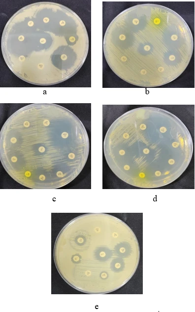
Figure 7. Antibiotics sensitivity of C. flaccumfaciens strains.a) SHGH1 strain b) SHGH2 strain c) SHGH3 strain d) SHGH4 strain e) SHGH5 strain
Molecular identification
The 16S rRNA gene is used for genetics analysis because it is highly conserved across many species of bacteria. The 16S rRNA full region of C. flaccumfaciens was amplified using 27F and 1522R primers, see (Section 2.5). The running agarose gel was visualized by a UV transilluminator (figure 8). Retrieved sequences were blasted in NCBI database for homology search. The 16S rRNA sequences of SHGH1 (OQ799009), SHGH2 (OQ7990010), SHGH3 (OQ799011), SHGH4 (OQ799012), and SHGH5 (OR230004) strains showed a homology of (99.49%, 97.36%, 99.20%, 99.00%, and 99.32%,) with deposited sequences in NCBI (Accessions: KP898898, MT323132, KT614051, MK389451, and MN826580 ), respectively.
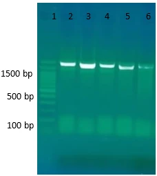
Figure 8. Amplified 16S rRNA region of C. flaccumfaciens. The Agarose gel image shows: Lane (1) 100 bp DNA ladder, lanes (2-6) 16S rRNA gene product amplified for SHGH1, SHGH2, SHGH3, SHGH4, and SHGH5 strains, respectively. Fragment size is 1495 bp
Phenotypic diagnosis of the colonies was carried out using the color, size, shape, and edge characteristics. The colonies had a yellow to orange color, smooth, shiny, sharp edges. These results are consistent with [9], [35], [36]. Biochemical tests included gram stain test, oxidase activity, catalase activity, motility test, and antibiotic susceptibility test. The bacterial cells were positive for the gram stain test, and were stained in a blue or purple color (figure 3). This might be as a result of peptidoglycan being present in the cell walls of gram-positive bacteria [10], [37]. Our results concur well with [7], [38]. The oxidase test was negative with cultured C. flaccumfaciens strains, as there was no color changing to purple (figure 4). This may mean that these bacteria do not have the cytochrome c oxidase that oxidizes the test reagent (Hederstedt, 2022 ), [39]. This result matches with [7], [10], [40]. Moreover, the result of the catalase test was positive, as the air bubbles were observed on the slide (figure 5), it means that these bacteria are able to produce catalase enzymes. The catalase enzyme is synthesized by aerobic bacteria, to protect from the toxic or oxidative damage of hydrogen peroxide of secondary products of oxygen metabolism [41], [42]. These findings are consistent with [7], [9], [28]. Furthermore, a semi-solid medium inoculated with C. flaccumfaciens was used to test motility. The result revealed that the bacteria growth had spread beyond the inoculation line (figure 6). It seems likely that this bacteria is motile. The motile bacteria actually produce a diffuse, spreading growth which is visible to the naked eye [43]. Our results are in line with [7], [10]. In addition, the sensitivity of C. flaccumfaciens for antibiotics was determined using the agar disc diffusion method. The C. flaccumfaciens sensitivity for antibiotics has been determined using the agar disc diffusion method. The results highlighted that all strains were sensitive to Meropenem, Chloramphenicol, Gentamicin, Amikacin, Ciprofloxacin, Nitrofurantoin, and Trimethoprim, while they were resistant to Cefixime and Cefotaxime (figure 7). The stronger antibiotic was Meropenem, where the growth inhibition zone was (2.8, 3.6, 3.5, 2.0, 2.0 mm), then Ciprofloxacin (2.8, 2.6, 3.4, 2.8, 0.9 mm). It could be because the gram-positive bacterium lacks the lipopolysaccharides layer found in Gram-negative bacteria [44], [45]. This makes it easier for cell-wall active antibiotics to reach their target (peptidoglycan layer). However, the reasons may be why the gram-positive bacteria develop the resistance to antibiotics (Cefixime and Cefotaxime ) are either by production of β-lactamases, an enzyme that breaks down antibiotics, or by altering the native penicillin-binding protein genes to reduce the affinity and sensitivity of the desired site, the (PBP) [46], [47]. The molecular analysis was done through the 16S rRNA region, a highly conserved sequence in bacteria. The result showed a homology of 99.49%, 97.36%, 99.20%, 99.00%, and 99.32% for the strains SHGH1, SHGH2, SHGH3, SHGH4, and SHGH5 (OR230004) with the submitted sequences in GenBank (Accessions: KP898898, MT323132, KT614051, MK389451, and MN826580), respectively, see (Figures 9-13). Orthologs, paralogs, and xenologs are the possible explanation and the reasons for shared ancestral sequences that resulted via speciation, duplication, or horizontal gene transfer, respectively [48].
C. flaccumfaciens is yellow-orange in color, a short rod shape, smooth, and shiny with a sharp edge. C. flaccumfaciens is a gram-positive bacterium, motile, oxidase-negative, catalase-positive, sensitive to Amikacin, Ciprofloxacin, Trimethoprim, Meropenem, Chloramphenicol, Gen-tamicin, and Nitrofurantoin antibiotics, resistance to Cefixime and Cefotaxime antibiotics. The 16S rRNA sequences of SHGH1, SHGH2, SHGH3, SHGH4, and SHGH5 strains were found to be a homology of 99.49%, 97.36%, 99.20%, 99.00%, and 99.32% with deposited sequences in NCBI (Accessions: KP898898, MT323132, KT614051, MK389451, and MN826580 ), respectively.
1 A.C. Parte, International journal of systematic and evolutionary microbiology 68(6) (2018) 1825. https://doi.org/10.1099/ijsem.0.002786
2 E. Osdaghi, A.J. Young, R.M. Harveson, Molecular plant pathology 21(5) (2020) 605. https://doi.org/10.1111/mpp.12926
3 Z. Cui, B. Steven, Q. Zeng, Microbiology Resource Announcements 10(19) (2021) 10. https://doi.org/10.1128/mra.00154-21
4 S. Seaton, J. Lemaire, P. Inderbitzin, V. Knight-Connoni, J.F. White, M.E. Trujillo, bioRxiv (2021). https://doi.org/10.1101/2021.03.18.434777
5 Y. Wei, X. Gao, Microbiology Resource Announcements 10(10) (2021) 10. 6. P.M. Guimar aes, J.J. Smith, S. Palmano, and G.S. Saddler, European Journal of Plant Pathology 109 (2003) 817 https://doi.org/10.1128/mra.01467-20
6 P.M. Guimar aes, J.J. Smith, S. Palmano, and G.S. Saddler, European Journal of Plant Pathology 109 (2003) 817. https://doi.org/10.1023/A:1026197914417
7 Y. Tumbarski, V. Georgiev, R. Nikolova, and A. Pavlov, The Journal of Microbiology, Biotechnology and Food Sciences 7(6) (2018) 623. http://dx.doi.org/10.15414/jmbfs.2018.7.6.623-627
8 R.M. Harveson, H.F. Schwartz, C.A. Urrea, and C.D. Yonts, Plant Disease 99(12) (2015) 1665. https://doi.org/10.1094/PDIS-03-15-0299-FE
9 E. Osdaghi, S.M. Taghavi, H. Hamzehzarghani, A. Fazliarab, R.M. Harveson, and J. R. Lamichhane, European Journal of Plant Pathology 146 (2016) 129. http://dx.doi.org/10.1007/s10658-016-0900-3
10 Y.F. Chen, Y.N. Yin, X.M. Zhang, and J.H. Guo, Plant Disease 91(6) (2007) 677. https://doi.org/10.1094/PDIS-91-6-0677
11 H.C. Huang, R.S. Erickson, P.M. Balasubramanian, T.F. Hsieh, and R.L. Conner, Canadian Journal of Plant Pathology 31(3) (2009) 290. http://dx.doi.org/10.1080/07060660909507603
12 E. Osdaghi, B. Pakdaman Sardrood, M. Bavi, N. Akbari Oghaz, S. Kimiaei, and S. Hadian, Journal of Phytopathology 163(7-8) (2015) 653. http://dx.doi.org/10.1111/jph.12300
13 B.A. Wood, and W.J. Easdown, Australasian Plant Pathology 19(1) (1990) 16. https://doi.org/10.1071/APP9900016
14 E. Osdaghi, S.M. Taghavi, A. Fazliarab, E. Elahifard, and J.R. Lamichhane, Crop Protection 78 (2015) 185. http://dx.doi.org/10.1016/j.cropro.2015.09.015
15 R.L. Conner, P. Balasubramanian, R.S. Erickson, H.C. Huang, and H.H. Mündel, Canadian Journal of Plant Science 88(6) (2008) 1109. https://doi.org/10.4141/CJPS08074
16 I.V. Agarkova, P.A. Lambrecht, A.K. Vidaver, and R.M. Harveson, Canadian journal of microbiology 58(6) (2012) 788. https://doi.org/10.1139/w2012-052
17 F. Schluenzen, A. Tocilj, R. Zarivach, J. Harms, M. Gluehmann, D. Janell, A. Bashan, H. Bartels, I. Agmon, F. Franceschi, A. Yonath, Cell 102(5) (2000) 615. https://doi.org/10.1016/s0092-8674(00)00084-2
18 T. Coenye, and P. Vandamme, FEMS microbiology letters 228(1) (2003) 45. https://doi.org/10.1016/s0378-1097(03)00717-1
19 M. Tsukuda, K. Kitahara, and K. Miyazaki, Scientific reports 7(1) (2017) 9993. https://doi.org/10.1038/s41598-017-10214-3
20 F. Pereira, J. Carneiro, R. Matthiesen, B. van Asch, N. Pinto, L. Gusmao, and A. Amorim, Nucleic acids research 38(22) (2010) e203. https://doi.org/10.1093%2Fnar%2Fgkq865
21 J.E. Clarridge III, Clinical microbiology reviews 17(4) (2004) 840. https://doi.org/10.1128/cmr.17.4.840-862.2004
22 M. Moriondo, L. Leolini, L. Brilli, C. Dibari, R. Tognetti, A. Giovannelli, B. Rapi, P. Battista, G. Caruso, R. Gucci, G. Argenti, A. Raschi, M. Centritto, C. Cantini, M. Bindi European Journal of Agronomy 105 (2019) 129. https://doi.org/10.1016/j.eja.2019.02.002
23 R. Marasco, M. Fusi, E. Rolli, B. Ettoumi, F. Tambone, S. Borin, H.-I. Ouzari, A. Boudabous, C. Sorlini, A. Cherif, F. Adani, D. Daffonchio, Environmental Microbiology 23(10) (2021) 6275. https://doi.org/10.1111/1462-2920.15764
24 M. Cheffi, A. Chenari Bouket, F.N. Alenezi, L. Luptakova, M. Belka, A. Vallat, M.E. Rateb, S. Tounsi, M. Ali Triki, L. Belbahri, Microorganisms 7(9) (2019) 314. https://doi.org/10.3390/microorganisms7090314
25 A. Afkhamifar, C. Moslemkhani, N. Hasanzadeh, and J. Razmi, European Journal of Plant Pathology 165(4) (2023) 611. http://dx.doi.org/10.1007/s10658-022-02631-6
26 C. Yazigi, M.S. Chaar, R. Busch, and M. Kern, Materials 16(15) (2023) 5305. https://doi.org/10.3390%2Fma16155305
27 G.B. Morris, E.J. Ridgway, and S.K. Suvarna, Bancroft's Theory and Practice of Histological Techniques (2019) 254. https://doi.org/10.1016/B978-0-7020-6864-5.00016-5
28 R. Nofiani, R. Rizky, R. Briliantoro, and P. Ardiningsih, Biodiversitas Journal of Biological Diversity 23(1) (2022) 453.
29 P. Shields, and L. Cathcart, Motility test medium protocol, American society for microbiology (2010).
30 Y. Jin, H. Xu, Q. Yao, B. Gu, Z. Wang, T. Wang, X. Yu, Y. Lu, B. Zheng, Y. Zhang, Frontiers in Microbiology 12 (2022) 780183. https://doi.org/10.3389%2Ffmicb.2021.780183
31 P. Shields, and L. Cathcart, Oxidase test protocol, American Society for Microbiology (2010).
32 R. Tarakanov, B. Shagdarova, T. Lyalina, Y. Zhuikova, A. Il’ina, F. Dzhalilov, V. Varlamov, Polymers 15(5) (2023) 1100.
33 B. Berbers, A. Saltykova, C. Garcia-Graells, P. Philipp, F. Arella, K. Marchal, R. Finand, K. Vanneste, N.H.C. Roosens, S.C.J. De Keersmaecker, Scientific reports 10(1) (2020) 4310. https://doi.org/10.1038/s41598-020-61158-0
34 A.M. Khaleel, R.M. Faisal, and H.A. Altaii, Malaysian Journal of Microbiology 19(2) (2023) 115. http://dx.doi.org/10.21161/mjm.220105
35 A. Díez-Méndez, and R. Rivas, AIMS microbiology 3(3) (2017) 354. https://doi.org/10.3934%2Fmicrobiol.2017.3.354
36 P. Evseev, A. Lukianova, R. Tarakanov, A. Tokmakova, M. Shneider, A. Ignatov, K. Miroshnikov, Current Issues in Molecular Biology 44(2) (2022) 889. https://doi.org/10.3390%2Fcimb44020060
37 F. Jiang, C. Cai, L. Gao, X. Su, and S. Han, ACS omega 8(2) (2022) 2485. https://doi.org/10.1021/acsomega.2c06964
38 R.M. Soares, G.G.P. Fantinato, L.M. Darben, F.C. Marcelino-Guimarães, C.D.S. Seixas, and G.E.D.S. Carneiro, Tropical Plant Pathology 38(5) (2013) 452. https://doi.org/10.1590/S1982-56762013000500012
39 A. Shimada, T. Tsukihara, and S. Yoshikawa, Frontiers in Chemistry 11 (2023) 1108190. https://doi.org/10.3389/fchem.2023.1108190
40 EPPO, Curtobacterium flaccumfaciens pv. Flaccumfaciens, EPPO Bull. 41 (2011) 320.
41 A.U. Prastujati, M. Hilmi, A. Khusna, I.I. Arief, S. Makmur, and Q. Maulida, In IOP Conference Series: Earth and Environmental Science 1020(1) (2022) 012026. https://doi.org/10.1088/1755-1315/1020/1/012026
42 M.A. Chandra, Journal of Health Management and Pharmacy Exploration 1(2) (2023). https://doi.org/10.52465/johmpe.v1i2.152
43 E.F. Mayer, M.Sc. thesis, University of Toronto (2019) 119p.
44 K. Klobucar, and E.D. Brown, Current Opinion in Chemical Biology 66 (2022) 102099. https://doi.org/10.1016/j.cbpa.2021.102099
45 C.M. Wesseling, T.M. Wood, C.J. Slingerland, K. Bertheussen, S. Lok, and N.I. Martin, Molecules 26(7) (2021) 1954. https://doi.org/10.3390/molecules26071954
46 B. Berger-Bächi, International Journal of Medical Microbiology 292(1) (2022) 27. https://doi.org/10.1078/1438-4221-00185
47 J.M. Munita, A.S. Bayer, and C.A. Arias, Evolving resistance among Gram-positive pathogens, Clinical Infectious Disease 61(2) (2015) S48. https://doi.org/10.1093/cid/civ523
48 E.V. Koonin, Annual Review of Genetics 39 (2005) 309. https://doi.org/10.1146/annurev.genet.39.073003.114725
Sh. Yaseen Taha, G.Q. Hasan, Morphological, biochemical, and molecular identification of Curtobacterium flaccumfaciens isolated from Olea europaea trees, UNEC J. Eng. Appl. Sci. 4(1) (2024) 83-91 https://doi.org/10.61640/ujeas.2024.0510
Anyone you share the following link with will be able to read this content:
This article is licensed under the Creative Commons Attribution ( CC BY 4.0 ) License, which permits unrestricted use, distribution, and reproduction in any medium, provided the original author and source are credited.


A.C. Parte, International journal of systematic and evolutionary microbiology 68(6) (2018) 1825. https://doi.org/10.1099/ijsem.0.002786
E. Osdaghi, A.J. Young, R.M. Harveson, Molecular plant pathology 21(5) (2020) 605. https://doi.org/10.1111/mpp.12926
Z. Cui, B. Steven, Q. Zeng, Microbiology Resource Announcements 10(19) (2021) 10. https://doi.org/10.1128/mra.00154-21
S. Seaton, J. Lemaire, P. Inderbitzin, V. Knight-Connoni, J.F. White, M.E. Trujillo, bioRxiv (2021). https://doi.org/10.1101/2021.03.18.434777
Y. Wei, X. Gao, Microbiology Resource Announcements 10(10) (2021) 10. 6. P.M. Guimar aes, J.J. Smith, S. Palmano, and G.S. Saddler, European Journal of Plant Pathology 109 (2003) 817 https://doi.org/10.1128/mra.01467-20
P.M. Guimar aes, J.J. Smith, S. Palmano, and G.S. Saddler, European Journal of Plant Pathology 109 (2003) 817. https://doi.org/10.1023/A:1026197914417
Y. Tumbarski, V. Georgiev, R. Nikolova, and A. Pavlov, The Journal of Microbiology, Biotechnology and Food Sciences 7(6) (2018) 623. http://dx.doi.org/10.15414/jmbfs.2018.7.6.623-627
R.M. Harveson, H.F. Schwartz, C.A. Urrea, and C.D. Yonts, Plant Disease 99(12) (2015) 1665. https://doi.org/10.1094/PDIS-03-15-0299-FE
E. Osdaghi, S.M. Taghavi, H. Hamzehzarghani, A. Fazliarab, R.M. Harveson, and J. R. Lamichhane, European Journal of Plant Pathology 146 (2016) 129. http://dx.doi.org/10.1007/s10658-016-0900-3
Y.F. Chen, Y.N. Yin, X.M. Zhang, and J.H. Guo, Plant Disease 91(6) (2007) 677. https://doi.org/10.1094/PDIS-91-6-0677
H.C. Huang, R.S. Erickson, P.M. Balasubramanian, T.F. Hsieh, and R.L. Conner, Canadian Journal of Plant Pathology 31(3) (2009) 290. http://dx.doi.org/10.1080/07060660909507603
E. Osdaghi, B. Pakdaman Sardrood, M. Bavi, N. Akbari Oghaz, S. Kimiaei, and S. Hadian, Journal of Phytopathology 163(7-8) (2015) 653. http://dx.doi.org/10.1111/jph.12300
B.A. Wood, and W.J. Easdown, Australasian Plant Pathology 19(1) (1990) 16. https://doi.org/10.1071/APP9900016
E. Osdaghi, S.M. Taghavi, A. Fazliarab, E. Elahifard, and J.R. Lamichhane, Crop Protection 78 (2015) 185. http://dx.doi.org/10.1016/j.cropro.2015.09.015
R.L. Conner, P. Balasubramanian, R.S. Erickson, H.C. Huang, and H.H. Mündel, Canadian Journal of Plant Science 88(6) (2008) 1109. https://doi.org/10.4141/CJPS08074
I.V. Agarkova, P.A. Lambrecht, A.K. Vidaver, and R.M. Harveson, Canadian journal of microbiology 58(6) (2012) 788. https://doi.org/10.1139/w2012-052
F. Schluenzen, A. Tocilj, R. Zarivach, J. Harms, M. Gluehmann, D. Janell, A. Bashan, H. Bartels, I. Agmon, F. Franceschi, A. Yonath, Cell 102(5) (2000) 615. https://doi.org/10.1016/s0092-8674(00)00084-2
T. Coenye, and P. Vandamme, FEMS microbiology letters 228(1) (2003) 45. https://doi.org/10.1016/s0378-1097(03)00717-1
M. Tsukuda, K. Kitahara, and K. Miyazaki, Scientific reports 7(1) (2017) 9993. https://doi.org/10.1038/s41598-017-10214-3
F. Pereira, J. Carneiro, R. Matthiesen, B. van Asch, N. Pinto, L. Gusmao, and A. Amorim, Nucleic acids research 38(22) (2010) e203. https://doi.org/10.1093%2Fnar%2Fgkq865
J.E. Clarridge III, Clinical microbiology reviews 17(4) (2004) 840. https://doi.org/10.1128/cmr.17.4.840-862.2004
M. Moriondo, L. Leolini, L. Brilli, C. Dibari, R. Tognetti, A. Giovannelli, B. Rapi, P. Battista, G. Caruso, R. Gucci, G. Argenti, A. Raschi, M. Centritto, C. Cantini, M. Bindi European Journal of Agronomy 105 (2019) 129. https://doi.org/10.1016/j.eja.2019.02.002
R. Marasco, M. Fusi, E. Rolli, B. Ettoumi, F. Tambone, S. Borin, H.-I. Ouzari, A. Boudabous, C. Sorlini, A. Cherif, F. Adani, D. Daffonchio, Environmental Microbiology 23(10) (2021) 6275. https://doi.org/10.1111/1462-2920.15764
M. Cheffi, A. Chenari Bouket, F.N. Alenezi, L. Luptakova, M. Belka, A. Vallat, M.E. Rateb, S. Tounsi, M. Ali Triki, L. Belbahri, Microorganisms 7(9) (2019) 314. https://doi.org/10.3390/microorganisms7090314
A. Afkhamifar, C. Moslemkhani, N. Hasanzadeh, and J. Razmi, European Journal of Plant Pathology 165(4) (2023) 611. http://dx.doi.org/10.1007/s10658-022-02631-6
C. Yazigi, M.S. Chaar, R. Busch, and M. Kern, Materials 16(15) (2023) 5305. https://doi.org/10.3390%2Fma16155305
G.B. Morris, E.J. Ridgway, and S.K. Suvarna, Bancroft's Theory and Practice of Histological Techniques (2019) 254. https://doi.org/10.1016/B978-0-7020-6864-5.00016-5
R. Nofiani, R. Rizky, R. Briliantoro, and P. Ardiningsih, Biodiversitas Journal of Biological Diversity 23(1) (2022) 453.
P. Shields, and L. Cathcart, Motility test medium protocol, American society for microbiology (2010).
Y. Jin, H. Xu, Q. Yao, B. Gu, Z. Wang, T. Wang, X. Yu, Y. Lu, B. Zheng, Y. Zhang, Frontiers in Microbiology 12 (2022) 780183. https://doi.org/10.3389%2Ffmicb.2021.780183
P. Shields, and L. Cathcart, Oxidase test protocol, American Society for Microbiology (2010).
R. Tarakanov, B. Shagdarova, T. Lyalina, Y. Zhuikova, A. Il’ina, F. Dzhalilov, V. Varlamov, Polymers 15(5) (2023) 1100.
B. Berbers, A. Saltykova, C. Garcia-Graells, P. Philipp, F. Arella, K. Marchal, R. Finand, K. Vanneste, N.H.C. Roosens, S.C.J. De Keersmaecker, Scientific reports 10(1) (2020) 4310. https://doi.org/10.1038/s41598-020-61158-0
A.M. Khaleel, R.M. Faisal, and H.A. Altaii, Malaysian Journal of Microbiology 19(2) (2023) 115. http://dx.doi.org/10.21161/mjm.220105
A. Díez-Méndez, and R. Rivas, AIMS microbiology 3(3) (2017) 354. https://doi.org/10.3934%2Fmicrobiol.2017.3.354
P. Evseev, A. Lukianova, R. Tarakanov, A. Tokmakova, M. Shneider, A. Ignatov, K. Miroshnikov, Current Issues in Molecular Biology 44(2) (2022) 889. https://doi.org/10.3390%2Fcimb44020060
F. Jiang, C. Cai, L. Gao, X. Su, and S. Han, ACS omega 8(2) (2022) 2485. https://doi.org/10.1021/acsomega.2c06964
R.M. Soares, G.G.P. Fantinato, L.M. Darben, F.C. Marcelino-Guimarães, C.D.S. Seixas, and G.E.D.S. Carneiro, Tropical Plant Pathology 38(5) (2013) 452. https://doi.org/10.1590/S1982-56762013000500012
A. Shimada, T. Tsukihara, and S. Yoshikawa, Frontiers in Chemistry 11 (2023) 1108190. https://doi.org/10.3389/fchem.2023.1108190
EPPO, Curtobacterium flaccumfaciens pv. Flaccumfaciens, EPPO Bull. 41 (2011) 320.
A.U. Prastujati, M. Hilmi, A. Khusna, I.I. Arief, S. Makmur, and Q. Maulida, In IOP Conference Series: Earth and Environmental Science 1020(1) (2022) 012026. https://doi.org/10.1088/1755-1315/1020/1/012026
M.A. Chandra, Journal of Health Management and Pharmacy Exploration 1(2) (2023). https://doi.org/10.52465/johmpe.v1i2.152
E.F. Mayer, M.Sc. thesis, University of Toronto (2019) 119p.
K. Klobucar, and E.D. Brown, Current Opinion in Chemical Biology 66 (2022) 102099. https://doi.org/10.1016/j.cbpa.2021.102099
C.M. Wesseling, T.M. Wood, C.J. Slingerland, K. Bertheussen, S. Lok, and N.I. Martin, Molecules 26(7) (2021) 1954. https://doi.org/10.3390/molecules26071954
B. Berger-Bächi, International Journal of Medical Microbiology 292(1) (2022) 27. https://doi.org/10.1078/1438-4221-00185
J.M. Munita, A.S. Bayer, and C.A. Arias, Evolving resistance among Gram-positive pathogens, Clinical Infectious Disease 61(2) (2015) S48. https://doi.org/10.1093/cid/civ523
E.V. Koonin, Annual Review of Genetics 39 (2005) 309. https://doi.org/10.1146/annurev.genet.39.073003.114725