UNEC Journal of Engineering and Applied Sciences Volume 3, No 1, pages 15-20 (2023) Cite this article, ![]() 2290 https://doi.org/10.61640/ujeas.2023.0503
2290 https://doi.org/10.61640/ujeas.2023.0503
In recent years, an investigation of mechanical and physical properties of carbon nanotubes formed as a result of tubular bending of the graphene layer are widely investigated by world researchers [1]. Very interesting physical prorperties with extremely mechanical strength made this materials as an important reserach object for electronics and other modern technologies. Therefore, the X-ray phase analysis of carbon nanotubes obtained by arc discharge method was carried out in the study [2]. Thus, after the discovery of carbon nanotubes, the mass production of multi-walled carbon nanotubes was started investigate widely [3]. In electronics, intense studies have been conducted in the direction of the application of single-walled carbon nanotubes. Using the Fowler-Nordheim model, their use has been studied in nanoemitters depending on the geometrical structure of nanotubes [8]. The obtained carbon nanotubes are classified into different types, single-walled, double-walled and multi-walled. Since a certain time and high temperature are required for obtaining nanotubes, some researchers have proposed various approximations for the accuracy of the mentioned classifications [9,10,11]. Carbon nanotubes are extremely light and have high electrical and thermal properties [12, 13].
Carbon nanotubes are low-dimensional nanostructures and differ from other structures due to their richness of mechanical, electrical [4-7], optical and magnetic properties [14-{ref16}].They also attract attention due to the uniqueness of their properties and wide range of applications. Different methods have been proposed for obtaining single-walled and multi-walled carbon nanotubes with different geometric shapes and diameters [17].
Methods of obtaining carbon nanotubes are quite expensive [18]. Aerosol [19], arc discharge [1, 20], laser ablation [1, 21], and chemical vapor deposition [22, 23] methods are the most commonly used methods for their acquisition. Carbon nanotubes are intensively applied in various fields of physics, chemistry, healthcare, and production [22, 24, 25]. Due to the emission properties of carbon nanotubes, they are used in electronic displays, X-ray sources, and luminescence sources [26-23]. In this regard, the application of nanotubes in electronic devices was investigated in detail in [30]. The synthesis of fiber-shaped carbon nanotubes at different temperatures was performed by the authors of [31].
We obtained carbon nanotubes among the various methods of obtaining carbon nanotubes using the arc discharge method [32], and functionalization was carried out with the help of carboxyl groups [10]. One of the important properties of carbon nanotubes is that they have metallic, semiconducting and dielectric properties depending on their chirality [24]. The structure, quality, and surface morphology of the carbon nanotube obtained by the arc discharge method were studied by the scanning electron microscopy method. The carbon nanotube which we received was studied in a Zeiss Crossbeam 540 scanning electron microscope. SEM analyses have suitableparameters for sizes from 0.8 nm (15kV) to 1.6 nm (1kV), 10-106 times magnification, 4pA - 20 nA probe voltage, and 2-133 Pa vacuum level. In addition, it is possible to perform 2D and 3D images of samples and elemental analysis. For this, a certain volume was taken from the carbon nanotube and viewed at room temperature on the sample plate of the electron microscope.
SEM images were taken within 24 hours. The structure and quality of the carbon nanotube obtained by the arc discharge method was studied using X-ray phase analysis. The X-ray phase analysis was carried out in the D2 Phaser diffractometer manufactured by the German "Bruker" company, with CuKα rays (λ=1.5406Å) in the 2θ=0.50÷800 angle range. Crystallographic, structural studies, calculations were performed on the basis of EVA and TOPAZ programs.
X-ray phase analysis is considered one of the important analysis methods for determining the chemical composition of the studied sample. As a result of this method of analysis, monochromatic light rays fall on the sample and secondary electron emission occurs as a result of the excitation of the sample. At this time, peaks with different energies are observed in the spectrum. X-ray phase analysis also allows obtaining information about chemical bonds. The energy level of electrons is determined by chemical bonds and their location in the valence band.One of the default of this method is the use of approximately 5 mg of sample for the experiment [24,30]. During the X-ray phase diffraction analysis of carbon nanotubes, works related to the uniqueness of the structure, properties of the tube, possess small size [8] and the electronic and geometric structure of the tubes were investigated [31-33].
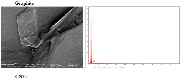
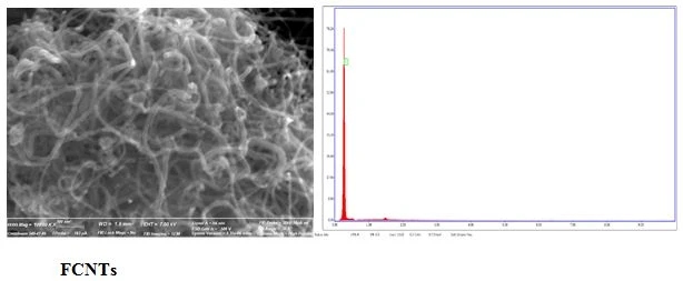
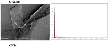
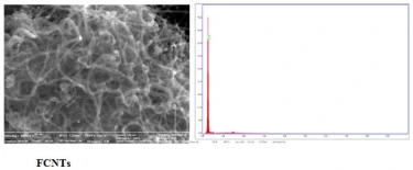
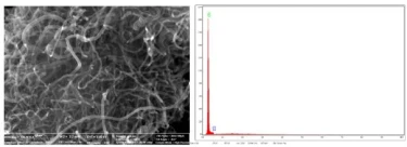
The purity, quality, surface morphology of graphite, carbon nanotube and functionalized carbon nanotube were investigated by scanning electron microscope (SEM) and energy dispersive analysis (EXD) (figure 1), and the structural properties were comparatively studied by X-ray phase analysis (figure 2).
SEM and EDX analysis has shown that high quality and high purity nanoscale materials under 100 nm diemnsions are obtained. Moreover, the structure of layered graphite is totally changed after functionalized by carboxyl group. More analytical experiments such as TEM or synchrotron are needed to get more precise and detailed information about the structure.
Graphite
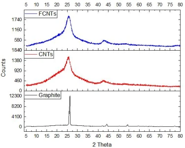
The graphite we use in our research has a crystalline structure of 99.9995% purity and Ultra "F" purity. X-ray phase analysis of graphite is shown in figure 2 and two sharp maxima were observed in the spectrum.
Figure 2. X-ray phase analyses of graphite, carbon nanotube, and carbon nanotube functionalized with a carboxyl group are shown, accordingly
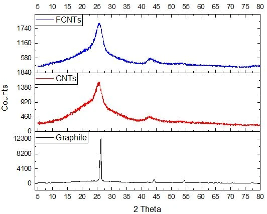
High quality graphite, carbon nanotube, and functionalized carbon nanotube by carboxyl group were synthesized using arc discharge method. SEM and EDX analysis has shown that the dimensions of the obtained materials are under 100 nm and applied carboxyl groups are infulenced on the structure of the graphite and made a significant changes. X-ray phas analysis has shown that synthesized graphite has crystalline structure with hexagonal symmetry and after functionalized by carboxyl groups, some amorphization is observed in the samples. However, more analytical investigations are needed to get more detailed information on this structures.
1 C. Journet, W.K. Maser, P. Bernier, A. Loiseau, M. Lamy de la Chapelle, S. Lefrant, P. Deniard, R. Lee, J.E. Fischer, Nature 388 (1997) 756.
2 X. Zhao, Y. Ando, Jpn.Jour.App.Phys. 37 (1998) 4846
3 D.T. Colbert, J. Zhang, S.M. Mcclure, P. Nikolaev, Z. Chen, J.H. Hafner, D.W. Owens, P.G. Kotula, C.B. Carter, J.H. Weaver, A.G. Rinzler, R.E. Smalley, Science 266 (1994) 1218.
4 R.G. Abaszade, Ecoenergetics 4 (2022) 46.
5 R.G. Abaszade, Ecoenergetics 4 (2022) 3.
6 A.G. Mammadov, R.G. Abaszade, V.O. Kotsyubynsky, E.Y. Gur, I.Y. Bayramov, E.A. Khanmamadova, O.A. Kapush, International Journal on Technical and Physical Problems of Engineering 14(3) (2022) 155.
7 Q.H. Wang, A.A. Setlur, J.M. Lauerhaas, J.Y. Dai, E.W. Seelig, R.P.H. Chang, Appl.Phys.Lett. 72 (1998) 2912.
8 S. Suzuki, C. Bower, Y. Watanabe, O. Zhou, Appl.Phys.Lett.76 (2000) 4007.
9 F.G. Brunetti, M.A. Herrero, J.D.M. Munoz, S. Giordani, A. Diaz-Ortiz, S. Filippone, G. Ruaro, M. Mneghetti, M. Prato, E. Vazquez, J.Am.Chem.Soc. 129 (2007) 14580.
10 M. Holzinger, J. Abraham, P. Whelan, R. Graupner, L. Ley, F. Hennrich, M. Kappes, A. Hirsch, Jour.Ame.Chem.Soc. 125 (2003) 8566.
11 H. Hu, B. Zhao, M.A. Hamon, K. Kamaras, M.E. Itkis, R.C. Haddon, Jour.Ame. Chem.Soc. 125 (2003) 14893.
12 T. Halicioglu, Thin Solid Films 312 (1998) 11.
13 E.I. Waldorff, A.M. Wass, P.P. Friedmann, M. Keidar, Jour.App.Phys. 95(5) (2004) 2749.
14 M.S. Dresselhaus, G. Dresselhaus, A. Jorio, Annual Review of Materials Research 34 (2004) 247.
15 T.P. Dyachkova, A.G. Tkachev, Methods of functionalization and modification of carbon nanotubes, Moscow: Spektr Publishing House (2013) 152 p.
16 M.M. Shokrieh, R. Rafiee, Materials 46(2) (2010) 155.
17 K. Awasthi, A. Srivastava, O.N. Srivastava, Journal of nanoscience and nanotechnology, 5 (2005) 1616.
18 W.H. Tan, S.L. Lee, J-H. Ng, W.W.F. Chong, C.T. Chong, I.J.Tech.7(2) (2016) 343.
19 A.G. Nasibulin, A. Moisala, D.P. Brown, H. Jiang, E.I. Kauppinen, Chemical Physics Letters 402 (2005) 227.
20 S. Iijima, Nature 354 (1991) 56.
21 W. Krätschmer, L.D. Lamb, K. Fostiropoulos, D.R. Huffman, Nature 347 (1990) 354.
22 J. Kong, A.M. Cassell, H.J. Dai, Chem.Phys.Lett. 292 (1998) 567.
23 A.V. Makunin, N.G. Chechenin, Synthesis and properties of nanocarbon structures: textbook, M.: Universitetskaya kniga (2011) 150 p.
24 W.T. Ebbesen, Carbon nanotubes: Preparation and properties, 1st edition, Florida: CRC Press (1996) 304 p.
25 M.J. O’Connell, Carbon nanotubes: Properties and applications, 1st edition, CRC Press (2006) 360 p.
26 A.V. Eletskii, Phys.Usp. 40(9) (1997) 899.
27 E.G. Rakov, Russ.Chem.Rev 70(10) (2001) 827.
28 A.V. Eletskii, Phys.Usp. 45(4) (2002) 369.
29 F. Xu , X. Liu, D.T. Stephen, Carbon 44(3) (2006) 570.
30 Y. Ando, X. Zhao, New diamond and frontier carbon technology 16(3) (2006) 122.
31 Y. Akai, S. Saito, Physica E 29 (2005) 555.
32 R. Das, Sh. Hamid, Md. Ali, S. Ramakrishna, Y. Wu, Current Nanoscience 11(1) (2015) 1.
33 R.G. Abaszade, M.B. Babanli, V.O. Kotsyubynsky, A.G. Mammadov, E. Gür, О.А. Kapush, M.O. Stetsenko, R.I. Zapukhlyak, Physics and Chemistry of Solid State 24(1) (2023) 153.
M.O. Stetsenko, R.G. Abaszade, X-ray phase analysis of carbon nanotubes obtained by the arc dischargemethod, UNEC J. Eng. Appl. Sci. 3(1) (2023) 15-20 https://doi.org/10.61640/ujeas.2023.0503
Anyone you share the following link with will be able to read this content:
This article is licensed under the Creative Commons Attribution ( CC BY 4.0 ) License, which permits unrestricted use, distribution, and reproduction in any medium, provided the original author and source are credited.
C. Journet, W.K. Maser, P. Bernier, A. Loiseau, M. Lamy de la Chapelle, S. Lefrant, P. Deniard, R. Lee, J.E. Fischer, Nature 388 (1997) 756.
X. Zhao, Y. Ando, Jpn.Jour.App.Phys. 37 (1998) 4846
D.T. Colbert, J. Zhang, S.M. Mcclure, P. Nikolaev, Z. Chen, J.H. Hafner, D.W. Owens, P.G. Kotula, C.B. Carter, J.H. Weaver, A.G. Rinzler, R.E. Smalley, Science 266 (1994) 1218.
R.G. Abaszade, Ecoenergetics 4 (2022) 46.
R.G. Abaszade, Ecoenergetics 4 (2022) 3.
A.G. Mammadov, R.G. Abaszade, V.O. Kotsyubynsky, E.Y. Gur, I.Y. Bayramov, E.A. Khanmamadova, O.A. Kapush, International Journal on Technical and Physical Problems of Engineering 14(3) (2022) 155.
Q.H. Wang, A.A. Setlur, J.M. Lauerhaas, J.Y. Dai, E.W. Seelig, R.P.H. Chang, Appl.Phys.Lett. 72 (1998) 2912.
S. Suzuki, C. Bower, Y. Watanabe, O. Zhou, Appl.Phys.Lett.76 (2000) 4007.
F.G. Brunetti, M.A. Herrero, J.D.M. Munoz, S. Giordani, A. Diaz-Ortiz, S. Filippone, G. Ruaro, M. Mneghetti, M. Prato, E. Vazquez, J.Am.Chem.Soc. 129 (2007) 14580.
M. Holzinger, J. Abraham, P. Whelan, R. Graupner, L. Ley, F. Hennrich, M. Kappes, A. Hirsch, Jour.Ame.Chem.Soc. 125 (2003) 8566.
H. Hu, B. Zhao, M.A. Hamon, K. Kamaras, M.E. Itkis, R.C. Haddon, Jour.Ame. Chem.Soc. 125 (2003) 14893.
T. Halicioglu, Thin Solid Films 312 (1998) 11.
E.I. Waldorff, A.M. Wass, P.P. Friedmann, M. Keidar, Jour.App.Phys. 95(5) (2004) 2749.
M.S. Dresselhaus, G. Dresselhaus, A. Jorio, Annual Review of Materials Research 34 (2004) 247.
T.P. Dyachkova, A.G. Tkachev, Methods of functionalization and modification of carbon nanotubes, Moscow: Spektr Publishing House (2013) 152 p.
M.M. Shokrieh, R. Rafiee, Materials 46(2) (2010) 155.
K. Awasthi, A. Srivastava, O.N. Srivastava, Journal of nanoscience and nanotechnology, 5 (2005) 1616.
W.H. Tan, S.L. Lee, J-H. Ng, W.W.F. Chong, C.T. Chong, I.J.Tech.7(2) (2016) 343.
A.G. Nasibulin, A. Moisala, D.P. Brown, H. Jiang, E.I. Kauppinen, Chemical Physics Letters 402 (2005) 227.
S. Iijima, Nature 354 (1991) 56.
W. Krätschmer, L.D. Lamb, K. Fostiropoulos, D.R. Huffman, Nature 347 (1990) 354.
J. Kong, A.M. Cassell, H.J. Dai, Chem.Phys.Lett. 292 (1998) 567.
A.V. Makunin, N.G. Chechenin, Synthesis and properties of nanocarbon structures: textbook, M.: Universitetskaya kniga (2011) 150 p.
W.T. Ebbesen, Carbon nanotubes: Preparation and properties, 1st edition, Florida: CRC Press (1996) 304 p.
M.J. O’Connell, Carbon nanotubes: Properties and applications, 1st edition, CRC Press (2006) 360 p.
A.V. Eletskii, Phys.Usp. 40(9) (1997) 899.
E.G. Rakov, Russ.Chem.Rev 70(10) (2001) 827.
A.V. Eletskii, Phys.Usp. 45(4) (2002) 369.
F. Xu , X. Liu, D.T. Stephen, Carbon 44(3) (2006) 570.
Y. Ando, X. Zhao, New diamond and frontier carbon technology 16(3) (2006) 122.
Y. Akai, S. Saito, Physica E 29 (2005) 555.
R. Das, Sh. Hamid, Md. Ali, S. Ramakrishna, Y. Wu, Current Nanoscience 11(1) (2015) 1.
R.G. Abaszade, M.B. Babanli, V.O. Kotsyubynsky, A.G. Mammadov, E. Gür, О.А. Kapush, M.O. Stetsenko, R.I. Zapukhlyak, Physics and Chemistry of Solid State 24(1) (2023) 153.