UNEC Journal of Engineering and Applied Sciences Volume 2 No 1, pages 85-91 (2022) Cite this article, ![]() 1923
1923
Recently, rare-earth doped semiconductor materials are widely investigated due to wide application area and high perpspectives [1-8]. In particular, since the 70s of last century, it is known that rare-earth doped alkaline earth sulphides are high promising phosphor materialsin terms of application capabilities in the various fields of industry and technology [9-15]. Currently, rare-earth doped alkaline earth thiogallates is of particular importance in the lighting technologies as a phosphors due to high emission intensity and efficiency properties [16-18]. Thus, only materials with appropriate emission and excitation parameters can be applied to obtain white LEDs. From this point of view, the application of the above-mentioned thiogallates in this direction have a great importance. High symmetry in crystallization, easy and cheap technogical process, high resistivity to temperature and humidity, chemical stability, as well as high emission intensity and efficiency increases application pespectives of these types of materials. Alkaline earth thiogallates can doped up to high concentration of rare-earth elements due to closer ionic radii. Therefore, rare-earth ions are easily substitute alkaline earth ions in the matrices and located in the nodes of crystals lattice.
In general, sufficiently high number of investigations have been devoted to study crystal structure of alkaline earth thiogallates [15, 19-23]. Depending on the alkaline earth elements (Ca, Ba, Sr) the host matrix have high symmetry cubic or orthorhombic structure. Also, changes in the crystal structures with the partial replacement of alkaline earth elements in the same matrix were investigated and the transition from the cubic phase to the orthorombic phase depending on the concentration and vice versa were studied in detail [15, 24, 25].
It is known that, the emission properties of the phosphors are different in samples depending on the rare-earth ions. In this regard, Eu2+ activated materials is of particular importance. Alkaline earth sulfides activated by the Eu2+ ion have a high potential for the formation of white light emitting diodes, both in terms of the required excitation source and the emission properties. As is known, there color of light (blue, green and red) are required to obtain white light. Blue diodes, which are commonly used to obtain white light and are also the excitation source for phosphorus. Green emission can be easily obtained from phosphors (eg, BaGa2S4:Eu, SrGa2S4:Eu, etc. [26-29]) obtained after activation of the above-mentioned thiogallates with rare earth elements. Here, the main problem is always the obtain red emission phosphors, which, in addition to red emission, these materials should be resistant to various influences (air, temperature, humidity, etc.), as well as have a high intensity and efficiency. Until today, several studies have studied several red phosphors, which are considered promising materials in terms of application [30-32].
Eu doped CaS compound is a good candidate as high intensity red emission phosphor, which emission and required excitation properties are appropriate to application in the white light technology. Although the some emission properties of this sample were studied by researchers [33, 34], but the structure and thermal properties of the materials have not been sufficiently studied. Therefore, in this study, the structure and the thermal properties of undoped and Eu2+ doped CaS compounds were investigated at room temperature and in the temperature range of 290-1273 K, respectively.
CaS and CaS:Eu were synthesized by different methods. Undoped CaS was synthesized from CaCO3 at H2S environment. The chemical reaction was carried out at 800 oC temperature during 24 h. H2S steam was obtained from decomposition of ammonium thiocyanate (NH4SCN) at 250 oC. Argon (Ar) inert gas was used to move the H2S steam in the system, as well as removing residual gases from the system. Eu doped CaS compounds was synthesized by solid state reaction at high temperature (1200 oC) and high vacuum (10-3-10-4 torr) in the quartz ampoule. Synthesis was carried out for 4 hours and then the temperature of the furnace down to 800 oC for annealing. After 5 hours of annealing process, the furnace turned off and cooled with ampoule.
X-ray diffraction measurements were performed at D8 ADVANCE (Bruker) powder diffractometer at room temperature. Thermal measurements were conducted using “Perkin Elmer” STA 6000 differential thermal analyzer. Operating temperature range was 20-1000 oC with 5, 10, 15 and 20 K/min. Polyscience analyzer and “digital temperature controller” was cooling system. Kinetic parameters were determined using “Pyris Manager” software. Argon inert gas injected into the system at a rate of 20 ml/min to remove the combustion products and prevent the condensation process. Standard aluminum oxide based pan (177.78 mg) was used for measurements. The weight of the sample was measured with 10-6 accuracy and automatically recorded. The software automatically determines the difference between the mass of the sample-filled pan and the empty pan. The specified mass is stored in the software. The parameters of endo and exothermic effects in thermal spectra are calculated using the Calculation menu. All experimental and theoretically calculated results are graphically described in the program "OriginPro 9.0".
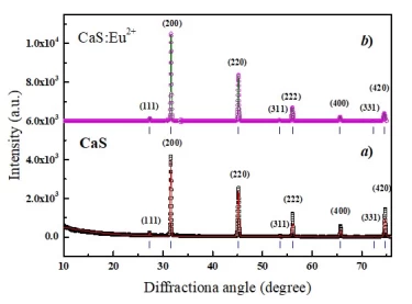
The crystal structure of CaS polycrystals were investigated by angle dispersive X-ray diffraction method. The measured spectra at normal condition is given in Figure 1a. As can be seen from the spectra, 8 diffraction peaks are observed in the range of 2θ = 10-75°. These maxima corresponds to (111), (200), (220), (311), (222), (400), (331) and (420) planes. X-ray diffraction spectra of CaS was analyzed using Rietveld method [35]. As a result of analysis, it was determined that the crystal structure of CaS compound has a high symmetry cubic phase with Fm m (225) space group. Lattice parameters of the structure is determined as: a = b = c = 5.6872 Å. Obtained result is consistent with the results of previous studies [36, 37].
Figure 1. Angle dispersive X-ray diffraction spectra of CaS (a) and CaS:Eu2+ (b) polyscrystals.
The crystal structure of EuF2 doped (3 mol%) CaS polycrystal also studied by angle dispersive X-ray diffraction method. Obtained result at normal condition is given in Figure 1b. Similarly to undoped CaS compound, 8 diffraction peaks are also observed for CaS:Eu2+ polycrystal in the range of 2θ = 10-75°. Observed peaks corresponds to (111), (200), (220), (311), (222), (400), (331) and (420) planes. It was determined that the crystal structure CaS:Eu2+ compound is high symmetry cubic phase with Fm m (225) space group. There is a slight difference with undoped CaS in the lattice parameters. Lattice parameters of the CaS:Eu2+ polycrystal are a = b = c = 5.6984 Å.
A comparison of the X-ray diffraction results have shown that the crystal structure of undoped and Eu2+ doped CaS compounds are compatible with each other. This is due to the EuF2 compound was added to CaS structure with small amount (3 mol%). In this case, divalent Ca2+ cations are partially substitute with Eu2+ ions. F atoms on the EuF2 separate in the form of gas (F2↑). There are not observed fundamental changes in the structure of host matrix due to low concentration of Eu2+ ions. The difference in the lattice parameters is due to the ionic radii of the binary metals Ca2+ and Eu2+. It is known that ionic radii of calcium atoms is RCa2+= 1.03 Å, and europium atoms is REu2+= 1.18Å. The difference between ionic radii of ΔR = 0.15 Å cause to difference between lattice parameters of Δa = 0.0112 Å. If Ca atoms are replaced more with Eu atoms, then the cubic symmetry of the space group Fm3 ̅m (225) will still be maintained. Because the structure of the EuS compound also corresponds to this crystal structure [37, 38].
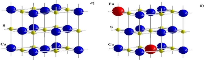
The crystal structure of the CaS compound is a model corresponding to the crystal structure of the NaCl compound [39]. Thus, divalent Ca2+ cation and S2- anion are located at the positions of monovalent Na1+ kation and Cl1- anion in crystal structure of NaCl and formed CaS crystal. In the crystal structure, calcium and sulfur atoms located at the coordinates of Ca (0, 0, 0) and S (0.5, 0.5, 0.5) alternate along the, and axes. The crystal structure of CaS compound is shown in Figure 2(a).
The crystal structure CaS:Eu2+ compound is somewhat more complex than that of the CaS compound. If the EuF2 crystals were suspended between the crystallites that forms the CaS polycrystals, then the crystal structure would be more complex. Although EuF2 crystals also correspond to the cubic symmetry of the space group Fm m (225), the concentration of F atoms and their positions in the crystal structure are different [40]. In our study, Eu2+ ions replaced Ca2+ ions in the crystal lattice, therefore a single-phase system was observed. However, it is probable that which Ca2+ ions replaced with Eu2+ ions. Therefore, these substitutions occur only as shown in Figure 2(b), affecting only the change in the lattice parameters.
Figure 2. Crsytal structures of CaS (a) and CaS:Eu2+ (b) compounds.
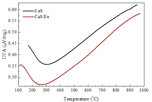
Differential Thermal Analysis (DTA) and Thermogravimetric Analysis (TG) of the CaS and CaS:Eu compounds were performed in the temperature range of 17-1000 oC. Examination of the DTA curves reveals that there is some slip in the spectra of the ordinate axis of the CaS and CaS: Eu crystals (Figure 3). This suggests that the Eu doped polycrystals consume less energy for the process to occur.
Figure 3. DTA spectra of CaS and CaS:Eu compounds.
There are observed endoeffecton the curves in both samples around a temperature of about 250 oC. Thus is most likely due to the removing or entry of water or any other mixtures in the system. Simultaneously, the observed endoeffects shift by about 50 oC relative to the temperature axis. This suggests that less energy is required in the thermodynamic system for these processes to occur when addition of Eu. However, more analytical work is needed to accurately confirm this.
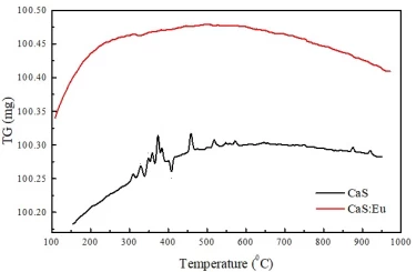
Moreover, TG analyzes of the sample were carried out in the same temperature range (Figure 4).
Figure 4. TG curves of CaS and CaS:Eu compounds.
It is clear from the TG analysis that the temperature increases in proportion to the temperature of the sample up to 250 oC. This growth is strongly observed in the Eu doped sample, which confirms the results of DTA analyzes. Thus, the increase in mass is an indication of the combination of any mixture from outside the system. Naturally, since less energy is required for the process to occurs in the Eu doped sample, the CaS:Eu sample absorbs foreign matter more rapidly. However, a decrease in the mass is observed for only Eu doped sample at temperatures above about 600 oC. It is assumed that the additional radicals are separated from the Eu doped sample at relatively high temperatures.
CaS and CaS:Eu compounds were synthesized by different methods. It was determined that observed 8 diffraction peaks corresponds to (111), (200), (220), (311), (222), (400), (331) and (420) planes. As a result of analysis by Rietveld method, it was determined that the crystal structure of CaS compound has a high symmetry cubic phase with Fm m (225) space group. Lattice parameters of the structure is determined as: a = b = c = 5.6872 Å. Crystal structure of Eu-doped CaS is similar to undoped CaS and it was determined that low concentration of Eu only influence to lattice parameters. Thus, the lattice parameters of CaS:Eu are a = b = c = 5.6984 Å. The probable location scheme of Eu atoms in the crystal lattice have been suggested. It was determined that endo effect is observed on the curves in both samples around a temperature of about 250 oC. Simultaneously, the observed endoeffects shift by about 50 oC relative to the temperature axis. This suggests that less energy is required in the thermodynamic system for these processes to occur when addition of Eu.
1 V. Hizhnyakov, V. Boltrushko, H. Kaasik, Y. Orlovskii, Optics Communications 485 (2021) 126693
2 N.L. Ignjatovic, L. Mančić, M. Vuković, Z. Stojanović, et al., Scientific Reports 9 (2019) 16305.
3 T. Zhong, J.M. Kindem, E.Miyazono and A. Faraon, Nature Communications 6(8206) (2015).
4 H.N. Van, D.D.T. Thuy, H.H. Van et al., New Journal of Chemistry 45(2) (2021) 751.
5 M.S. Leanenia, E.V. Lutsenko, M.V. Rzheutski, V.N. Pavlovskii, G.P. Yablonskii, T.G. Naghiyev, B.G. Tagiev, S.A. Abushev, O.B. Tagiev, Journal of Luminescence 181 (2017) 121.
6 D.T. Khan, N.T. Dang, S.H. Jabarov, T.G. Naghiyev, R.M. Rzayev, T.Q. Nguyen, H.V. Tuyen, N.T. Thanh, L.V.T. Son, Materials Research Express 7(1) (2020) 016507.
7 B.G. Tagiev, O.B. Tagiev, T.G. Nagiev, S.G. Asadullaeva, M.S. Leonenya, G.P.Yablonskii, S.A. Abushov, Optics and Spectroscopy 118(3) (2015) 389.
8 M.S. Leanenia, E.V. Lutsenko, M.V. Rzheutski, G.P. Yablonskii, T.G. Naghiyev, H.B. Ganbarova, O.B. Tagiev, Optical Materials 54 (2016) 45.
9 Sh.Tanaka, H. Yoshiyama, K. Nakamura, S. Wada et al., Jpn. J. Appl. Phys. 30(6A) (1991) L1021.
10 P.F. Smet, I. Moreels, Z. Hens, D. Poelman, Materials 3(4) (2010) 2834.
11 P. Benalloul, C. Barthou, C. Fouassier, A.N. Georgobiani, L.S. Lepnev, Y.N. Emirov, A. N. Gruzintsev, B.G. Tagiev, O.B. Tagiev, R.B. Jabbarov, Journal of The Electrochemical Society 150 (2003) 62.
12 R. Jabbarov, C. Chartier, B.G. Tagiev, O.B. Tagiev, N.N. Musayeva, C. Barthou, P. Benalloul, Journal of Physics and Chemistry of Solids 66 (2005) 1049.
13 M.S. Leanenya, E.V. Lutsenko, V.N. Pavlovskii, G.P.Yablonskii, T.G.Nagiev, B.G. Tagiev, O.B. Tagiev, S.A. Abushev, Journal of Applied Spectroscopy 82(1) (2015) 53.
14 M.S. Leanenia, E.V. Lutsenko, M.V. Rzheutski, V.N. Pavlovskii, G.P. Yablonskii, T.G. Naghiyev, B.G. Tagiev, S.A. Abushev, O.B. Tagiev, Doklady of the National Academy of Sciences of Belarus 59(6) (2016) 57.
15 B.G. Tagiyev, O.B. Tagiyev, A.I. Mammadov, Vu Xuan Quang, T.G. Naghiyev, S.H. Jabarov, M.S. Leonenya, G.P. Yablonskii, N.T. Dang, Physica B: Condensed Matter 478 (2015) 58.
16 Sh. Verma, K. Verma, D. Kumar, B. Chaudhary et al, Physica B: Condensed Matter 535 (2018) 106.
17 K. Korthout, P.F. Smet and D. Poelman, Appl. Phys. Lett. 98(26) (2011).
18 V. Jarý, L. Havlák, J. Bárta, M. Buryi et al, ECS Journal of Solid State Science and Technology 9(1) 2020.
19 T.E. Peters and J.A. Baglio, J. Electrochem. Soc.: Solid-State Scienceand Technology, 119(2) (1972) 230.
20 M. Al-Shakban, P.D. Matthews and P. O'Brien, Chemical Communications 72 (2017) 1.
21 C.Hidaka, T.Takizawa, Journal of Crystal Growth 237–239(3) (2002) 2009.
22 C. Komatsu-Hidaka, T. Takizawa, Journal of Crystal Growth, 222(3) (2001)574.
23 C. Hidaka, E. Yamagishi, T. Takizawa, Journal of Physics and Chemistry of Solids, 66(11) (2005) 2058.
24 H.S. Yoo, W.B. Im, S. Vaidyanathan, B.J. Park and D.Y. Jeon, Journal of The Electrochemical Society 155(3) (2008) J66-J70.
25 R. Yu, J. Wang, M. Zhang, H. Yuan, Journal of The Electrochemical Society 155(10) (2008) J290.
26 R.B. Jabbarov, C. Chartier, B.G. Tagiev, O.B. Tagiev, N.N. Musayeva, C. Barthou, P. Benalloul, 66(6) (2005) 1049.
27 A.N. Georgobiani, B.G. Tagiev, S.A. Abushov, O.B. Tagiev, Z. Xu, S. Zhao, Inorganic Materials 44 (2008) 110.
28 Z. Xinmin, W. Hao, Z. Heping, S. Qiang, Journal of Rare Earths 25(6) (2007) 701.
29 H. Kominami, Y. Nakanishi, K. Hara, Phys. Status Solidi C 12(6) (2015) 801.
30 J.Ch. Chang, Ch.T. Chen, M. Rudysh, M.G. Brikc, M. Piasecki, W.R. Liu, Journal of Luminescence 206 (2019) 417.
31 M. Rajendran and S. Vaidyanathan, New J. Chem., 44 (2020) 5354.
32 Z. Yang, Z. Yang, Q. Wei, Q. Zhou, Zh. Wang, Journal of Luminescence 210 (2019) 408.
33 S. Fukumoto, Y. Hayashi, S. Ibuki, K. Abe and H. Ohnishi, Jpn. J. Appl. Phys. 32 (1993) L791.
34 P.F. Smet, N. Avci, B. Loos, J.E. Van Haecke, and D. Poelman, J. Phys.: Condens. Matter 19 (2007) 246223.
35 J. Rodríguez-Carvajal, Physica B 192 (1993) 55.
36 I. Oftedal, Zeitschrift fuer Physikalische Chemie 128 (1927) 135.
37 O. Madelung, U. Rössler, M. Schulz, Springer-Verlag Berlin Heidelberg, 2000.
38 R.W.G. Wyckoff, Second edition. Interscience Publishers, New York, Crystal Structures 1 (1963) 467.
39 P. Juhás, Ch.L. Farrow, X. Yang, K.R. Knox, S.J.L. Billinge, Acta Crystallographica Section A 71 (2015) 562.
40 H. Grossholz, I. Hartenbach, G. Kotzyba, R. Pöttgen, Journal of Solid State Chemistry, 182(11) (2009) 3071.
T.G. Naghiyev, U.R. Rzayev, E.M. Huseynov, I.T. Huseynov, S.H. Jabarov, Effect of the europium doping on the calcium sulphide: structural and thermal aspects, UNEC J. Eng. Appl. Sci 2(1) (2022) 85-91
Anyone you share the following link with will be able to read this content:
This article is licensed under the Creative Commons Attribution ( CC BY 4.0 ) License, which permits unrestricted use, distribution, and reproduction in any medium, provided the original author and source are credited.


V. Hizhnyakov, V. Boltrushko, H. Kaasik, Y. Orlovskii, Optics Communications 485 (2021) 126693
N.L. Ignjatovic, L. Mančić, M. Vuković, Z. Stojanović, et al., Scientific Reports 9 (2019) 16305.
T. Zhong, J.M. Kindem, E.Miyazono and A. Faraon, Nature Communications 6(8206) (2015).
H.N. Van, D.D.T. Thuy, H.H. Van et al., New Journal of Chemistry 45(2) (2021) 751.
M.S. Leanenia, E.V. Lutsenko, M.V. Rzheutski, V.N. Pavlovskii, G.P. Yablonskii, T.G. Naghiyev, B.G. Tagiev, S.A. Abushev, O.B. Tagiev, Journal of Luminescence 181 (2017) 121.
D.T. Khan, N.T. Dang, S.H. Jabarov, T.G. Naghiyev, R.M. Rzayev, T.Q. Nguyen, H.V. Tuyen, N.T. Thanh, L.V.T. Son, Materials Research Express 7(1) (2020) 016507.
B.G. Tagiev, O.B. Tagiev, T.G. Nagiev, S.G. Asadullaeva, M.S. Leonenya, G.P.Yablonskii, S.A. Abushov, Optics and Spectroscopy 118(3) (2015) 389.
M.S. Leanenia, E.V. Lutsenko, M.V. Rzheutski, G.P. Yablonskii, T.G. Naghiyev, H.B. Ganbarova, O.B. Tagiev, Optical Materials 54 (2016) 45.
Sh.Tanaka, H. Yoshiyama, K. Nakamura, S. Wada et al., Jpn. J. Appl. Phys. 30(6A) (1991) L1021.
P.F. Smet, I. Moreels, Z. Hens, D. Poelman, Materials 3(4) (2010) 2834.
P. Benalloul, C. Barthou, C. Fouassier, A.N. Georgobiani, L.S. Lepnev, Y.N. Emirov, A. N. Gruzintsev, B.G. Tagiev, O.B. Tagiev, R.B. Jabbarov, Journal of The Electrochemical Society 150 (2003) 62.
R. Jabbarov, C. Chartier, B.G. Tagiev, O.B. Tagiev, N.N. Musayeva, C. Barthou, P. Benalloul, Journal of Physics and Chemistry of Solids 66 (2005) 1049.
M.S. Leanenya, E.V. Lutsenko, V.N. Pavlovskii, G.P.Yablonskii, T.G.Nagiev, B.G. Tagiev, O.B. Tagiev, S.A. Abushev, Journal of Applied Spectroscopy 82(1) (2015) 53.
M.S. Leanenia, E.V. Lutsenko, M.V. Rzheutski, V.N. Pavlovskii, G.P. Yablonskii, T.G. Naghiyev, B.G. Tagiev, S.A. Abushev, O.B. Tagiev, Doklady of the National Academy of Sciences of Belarus 59(6) (2016) 57.
B.G. Tagiyev, O.B. Tagiyev, A.I. Mammadov, Vu Xuan Quang, T.G. Naghiyev, S.H. Jabarov, M.S. Leonenya, G.P. Yablonskii, N.T. Dang, Physica B: Condensed Matter 478 (2015) 58.
Sh. Verma, K. Verma, D. Kumar, B. Chaudhary et al, Physica B: Condensed Matter 535 (2018) 106.
K. Korthout, P.F. Smet and D. Poelman, Appl. Phys. Lett. 98(26) (2011).
V. Jarý, L. Havlák, J. Bárta, M. Buryi et al, ECS Journal of Solid State Science and Technology 9(1) 2020.
T.E. Peters and J.A. Baglio, J. Electrochem. Soc.: Solid-State Scienceand Technology, 119(2) (1972) 230.
M. Al-Shakban, P.D. Matthews and P. O'Brien, Chemical Communications 72 (2017) 1.
C.Hidaka, T.Takizawa, Journal of Crystal Growth 237–239(3) (2002) 2009.
C. Komatsu-Hidaka, T. Takizawa, Journal of Crystal Growth, 222(3) (2001)574.
C. Hidaka, E. Yamagishi, T. Takizawa, Journal of Physics and Chemistry of Solids, 66(11) (2005) 2058.
H.S. Yoo, W.B. Im, S. Vaidyanathan, B.J. Park and D.Y. Jeon, Journal of The Electrochemical Society 155(3) (2008) J66-J70.
R. Yu, J. Wang, M. Zhang, H. Yuan, Journal of The Electrochemical Society 155(10) (2008) J290.
R.B. Jabbarov, C. Chartier, B.G. Tagiev, O.B. Tagiev, N.N. Musayeva, C. Barthou, P. Benalloul, 66(6) (2005) 1049.
A.N. Georgobiani, B.G. Tagiev, S.A. Abushov, O.B. Tagiev, Z. Xu, S. Zhao, Inorganic Materials 44 (2008) 110.
Z. Xinmin, W. Hao, Z. Heping, S. Qiang, Journal of Rare Earths 25(6) (2007) 701.
H. Kominami, Y. Nakanishi, K. Hara, Phys. Status Solidi C 12(6) (2015) 801.
J.Ch. Chang, Ch.T. Chen, M. Rudysh, M.G. Brikc, M. Piasecki, W.R. Liu, Journal of Luminescence 206 (2019) 417.
M. Rajendran and S. Vaidyanathan, New J. Chem., 44 (2020) 5354.
Z. Yang, Z. Yang, Q. Wei, Q. Zhou, Zh. Wang, Journal of Luminescence 210 (2019) 408.
S. Fukumoto, Y. Hayashi, S. Ibuki, K. Abe and H. Ohnishi, Jpn. J. Appl. Phys. 32 (1993) L791.
P.F. Smet, N. Avci, B. Loos, J.E. Van Haecke, and D. Poelman, J. Phys.: Condens. Matter 19 (2007) 246223.
J. Rodríguez-Carvajal, Physica B 192 (1993) 55.
I. Oftedal, Zeitschrift fuer Physikalische Chemie 128 (1927) 135.
O. Madelung, U. Rössler, M. Schulz, Springer-Verlag Berlin Heidelberg, 2000.
R.W.G. Wyckoff, Second edition. Interscience Publishers, New York, Crystal Structures 1 (1963) 467.
P. Juhás, Ch.L. Farrow, X. Yang, K.R. Knox, S.J.L. Billinge, Acta Crystallographica Section A 71 (2015) 562.
H. Grossholz, I. Hartenbach, G. Kotzyba, R. Pöttgen, Journal of Solid State Chemistry, 182(11) (2009) 3071.