UNEC Journal of Engineering and Applied Sciences Volume 1, No 1, pages 49-53 (2021) Cite this article, ![]() 1622
1622
The study of compounds with magnetic properties is one of the topical problems in condensed matter physics. It is known that some materials have both ferroelectric and ferromagnetic properties. Such compounds have more interesting physical properties. Barium hexaferrite and its solid solutions are considered to be such compounds. Determining the relationship between physical properties and structure of materials is very important when studying other physical properties of these components [1-3].
BaFe12O19, which has a hexagonal crystal structure, is one of the most studied multiferroics in recent years.Barium hexaferrite is a material with ferrimagnetic properties under normal conditions and at room temperature.It was found that the ferrimagnetic-paramagnetic phase transition in this compound occurs at a temperature TC ≈ 750 K. Previous structural studies have shown that when Fe3+ ions are replaced by Ga3+, Al3+, Sc3+ ions in barium hexaferrite, changes in the crystal structure of the compounds are observed [4-6]. An increase in the concentration of diamagnetic ions leads to a partial violation of the long-range magnetic order, thereby weakening the magnetic properties. Hexaferrites and composite materials based on them are widely used in various fields as materials that absorb electromagnetic radiation. Consequently, by preserving the magnetic properties, replacing Fe atoms with In changes the electronic configuration of these materials, making them important materials for electronics and spintronics.
It is known that the crystallite size also affects other properties of the compound.Therefore, it is important to study the size effects of each research object. In this work, the crystal structure, surface structure, and chemical analysis of the BaFe10.8In1.2O19 compound were carried out. The studies were performed at room temperature and under normal conditions by XRD and SEM.
2.1. Synthesis.
Investigated BaFe10.8In1.2O19 samples were obtained from high purity Fe2O3 and In2O3 oxides and carbonate BaCO3 by conventional solid reaction method using ‘two-steps’ topotactic reactions. At first the oxides and carbonate were mixed with design ratio. Then the prefiring was performed at 1200 ºC in air during 6 h. Final synthesis was carried out at 1300 ºC in air during 6 h. After synthesis the samples were slowly cooled (100 ºC h-1).
2.2. XRD analysis.
The X-ray diffraction measurement was performed using a Bruker D8 Advance powder diffractometer with the following parameters: 40 kV, 40 mA, Cu Kα radiation (λ = 1.5406Å). The X-ray diffraction data were treated using the FullProf program.
2.3. SEM (Scanning Electron Microscope) analysis.
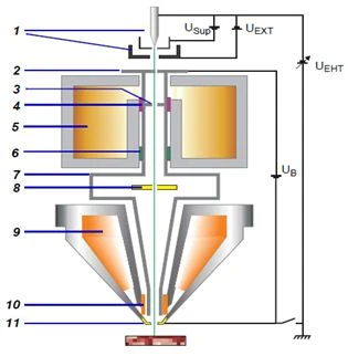
Surface morphology and chemical analysis of BaFe10.8In1.2O19 were performed on SEM (Scanning Electron Microscope, ZEISS, ΣIGMA VP). In a Scanning Electron Microscope, a tungsten element enclosed in a zirconium ring is used as an electronic source. The operating mode for the voltage during the thermal emission process based on the Schott effect is 0.05 V - 30 keV, the operating distance between the beam source and the sample is assumed to be ≤10 mm. Samples are prepared in specially selected laboratory conditions.
Figure 1. Schematic description of electron optics in Scanning Electron Microscopy: 1. Schottky field emission, 2. Anode, 3. Multichannel pores, 4. Grouping system, 5. Lens, 6. Stigmator, 7. Linear channel, 8. Detector, 9. Electromagnetic lenses, 10. Scanning camera, 11. Cover.
The carbon is pulled into trays and placed in silver slots. After placing the samples in the chamber, molecular pumps create a vacuum of 10-7 Mbar. The main purpose of creating a high vacuum is to increase the free path of electrons and reduce the likelihood of elastic or inelastic collisions.In this case, the energy transferred to the accelerated electrons is transferred directly to the interaction of the sample atoms, which leads to a decrease in the error.
Along with the experimental part, of greater interest is the theoretical part or optics of the processes occurring in electron optics under a microscope. A schematic sequence of processes in SEM is shown in detail in Figure 3. Under the action of thermal emission, electrons emitted from the tungsten element are directed by an electrostatic field. Accelerated electrons in a potential field are accelerated up to 30 kV and interact with the sample.
3.1. Structural analysis.
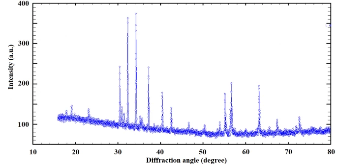
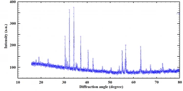
The crystal structure of BaFe10.8In1.2O19 was studied by X-ray diffraction. The X-ray diffraction spectrum obtained under normal conditions and at room temperature is shown in Figure 2.
Figure 2. X-ray diffraction spectrum of the compound Fe10.8In1.2O19.
As a result of the analysis of the X-ray diffraction spectrum obtained in the range of diffraction angles 15º ≤ 2θ ≤ 80º, it was determined that the crystal structure of the BaFe10.8In1.2O19 compound corresponds to the hexagonal symmetry of the space group P63/mmc.It is known that the crystal structure of barium hexaferrite also corresponds to the hexagonal symmetry of the space group P63/mmc [7-9]. As can be seen, no significant changes in the crystal structure are observed with the substitution of Fe → In up to a concentration of x = 1.2.
3.2. Surface structure.
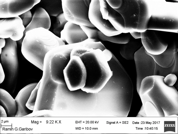
The surface structure of BaFe10.8In1.2O19was studied using a scanning electron microscope. A description of the surface structure is shown in Figure 3.
Figure 3. The surface structure of Fe10.8In1.2O19, obtained with a Scanning Electron Microscope.
As can be seen from Figure 3, the crystallites of the powder sample are of different sizes.The sizes of the smallest crystallites are d ~ 1 μm, and the sizes of the largest crystallites are d ~ 26 μm.From the morphology of the crystallites, it is clear that the sample was synthesized with high quality.The object of the study was not in the form of solid solutions (BaFe1.2O19–BaIn12O19), but was synthesized in a single-phase state - BaFe10.8In1.2O19.
3.3. Chemical analysis.
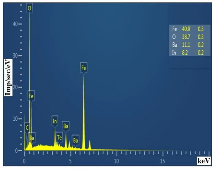
The chemical composition of BaFe10.8In1.2O19 was also studied during SEM analysis. The chemical analysis spectrum is shown in Figure 4.
Figura 4. Spectrum of analysis of the chemical composition of the BaFe10.8In1.2O19 compound.
As a result of the analysis of the composition, it was determined that BaFe10.8In1.2O19consists of 40.9% - Fe, 38.7% - O, 11.1% - Ba, and 8.2% - In. As you can see, the substance was synthesized with a fairly high purity. A sample was synthesized with a purity of 99.9%. In the composition, very small amounts of 0.1% of other elements were obtained, which were not observed in the X-ray diffraction spectrum.
The crystal structure and morphology of the BaFe10.8In1.2O19 compound were studied by X-ray diffraction and SEM. Chemical analysis of the samples was carried out. The crystal structure of the obtained compound is hexagonally symmetric with the space group P63 / mmc in accordance with the structures of barium hexaferrite and its solid solutions. When studying a powder sample by SEM, it was determined that the size of the smallest crystallites is d ~ 1 μm, and the size of the largest crystallites is d ~ 26 μm.
1 S.H. Jabarov, N.T. Dang, S.E. Kichanov, D.P. Kozlenko, L.S. Dubrovinsky, J.G. Park, S. Lee, A.I. Mammadov, R.Z. Mehdiyeva, B.N. Savenko, N.X. Nghia, L.H. Khiem, N.T.T. Lieu, L.T.P. Thao, Materials Research Express 6(8) (2019) 086110.
2 N.T. Dang, D.P. Kozlenko, N. Tran, B.W. Lee, T.L. Phan, R.P. Madhogaria, V. Kalappattil, D.S. Yang, S.E. Kichanov, E.V. Lukin, B.N. Savenko, P. Czarnecki, T.A. Tran, V.L. Vo, L.T.P. Thao, D.T. Khan, N.Q. Tuan, S.H. Jabarov, M.H. Phan, Journal of Alloys and Compounds 808 (2019) 151760.
3 D.P. Kozlenko, N.T. Dang, N.O. Golosova, S.E. Kichanov, E.V. Lukin, P.J.L. Kelley, E.M. Clements, K.V. Glazyrin, S.H. Jabarov, T.L. Phan, B.N. Savenko, H. Srikanth, M.H. Phan, Physical Review B 98(13) (2018) 13443.
4 A.I. Mammadov, N.T. Dang, R.Z. Mehdiyeva, A.V. Trukhanov, S.G. Asadullayeva, S.V. Trukhanov, R.E. Huseynov, S.H. Jabarov, Modern Physics Letters B, 34(35) (2020) 2050411.
5 A.I. Mammadov, N.T. Dang, R.Z. Mehdiyeva, À.V. Trukhanov, R.E. Huseynov, S.V. Trukhanov, S.H. Jabarov, Modern Physics Letters B 34(33) (2020) 2050381.
6 F.G. Agayev, S.H. Jabarov, G.Sh. Ayyubova, A.V. Trukhanov, S.V. Trukhanov, M.N. Mirzayev, T.G. Naghiyev, N.T. Dang, Journal of Superconductivity and Novel Magnetism 33 (2020) 2867-2873.
7 F.G. Agayev, S.H. Jabarov, G.Sh. Ayyubova, M.N. Mirzayev, S.V. Trukhanov, E.L. Trukhanova, M.A. Darwish, S.V. Podgornaya, D.A. Vinnik, T.P. Hoang, N.T. Dang, A.V. Trukhanov, Physica B: Condensed Matter 580 (2020) 411772.
8 R.E. Huseynov, A.I. Mammadov, R.Z. Mehdiyeva, A.V. Trukhanov, S.V. Trukhanov, V.A. Turchenko, T.P. Hoang, N.T. Dang, S.H. Jabarov, Journal of the Korean Physical Society 74(6) (2019) 584-588.
9 S.G. Dzhabarov, A.V. Trukhanov, S.V. Trukhanov, V.A. Turchenko, V.V. Oleinik, E.S. Yakovenko, L.Yu. Matsui, L.L. Vovchenko, V.L. LaunetsI, S. Kazakevich, Physics of the Solid State 58(9) (2016) 1792-1797.
G.Sh. Ayyubova, S.H. Jabarov, Crystal structure, surface structure and chemical analysis of BaFe10.8In1.2O19 compound. UNEC J. Eng. Appl. Sci. 1(1) (2021) 49-53
Anyone you share the following link with will be able to read this content:
This article is licensed under the Creative Commons Attribution ( CC BY 4.0 ) License, which permits unrestricted use, distribution, and reproduction in any medium, provided the original author and source are credited.


S.H. Jabarov, N.T. Dang, S.E. Kichanov, D.P. Kozlenko, L.S. Dubrovinsky, J.G. Park, S. Lee, A.I. Mammadov, R.Z. Mehdiyeva, B.N. Savenko, N.X. Nghia, L.H. Khiem, N.T.T. Lieu, L.T.P. Thao, Materials Research Express 6(8) (2019) 086110.
N.T. Dang, D.P. Kozlenko, N. Tran, B.W. Lee, T.L. Phan, R.P. Madhogaria, V. Kalappattil, D.S. Yang, S.E. Kichanov, E.V. Lukin, B.N. Savenko, P. Czarnecki, T.A. Tran, V.L. Vo, L.T.P. Thao, D.T. Khan, N.Q. Tuan, S.H. Jabarov, M.H. Phan, Journal of Alloys and Compounds 808 (2019) 151760.
D.P. Kozlenko, N.T. Dang, N.O. Golosova, S.E. Kichanov, E.V. Lukin, P.J.L. Kelley, E.M. Clements, K.V. Glazyrin, S.H. Jabarov, T.L. Phan, B.N. Savenko, H. Srikanth, M.H. Phan, Physical Review B 98(13) (2018) 13443.
A.I. Mammadov, N.T. Dang, R.Z. Mehdiyeva, A.V. Trukhanov, S.G. Asadullayeva, S.V. Trukhanov, R.E. Huseynov, S.H. Jabarov, Modern Physics Letters B, 34(35) (2020) 2050411.
A.I. Mammadov, N.T. Dang, R.Z. Mehdiyeva, À.V. Trukhanov, R.E. Huseynov, S.V. Trukhanov, S.H. Jabarov, Modern Physics Letters B 34(33) (2020) 2050381.
F.G. Agayev, S.H. Jabarov, G.Sh. Ayyubova, A.V. Trukhanov, S.V. Trukhanov, M.N. Mirzayev, T.G. Naghiyev, N.T. Dang, Journal of Superconductivity and Novel Magnetism 33 (2020) 2867-2873.
F.G. Agayev, S.H. Jabarov, G.Sh. Ayyubova, M.N. Mirzayev, S.V. Trukhanov, E.L. Trukhanova, M.A. Darwish, S.V. Podgornaya, D.A. Vinnik, T.P. Hoang, N.T. Dang, A.V. Trukhanov, Physica B: Condensed Matter 580 (2020) 411772.
R.E. Huseynov, A.I. Mammadov, R.Z. Mehdiyeva, A.V. Trukhanov, S.V. Trukhanov, V.A. Turchenko, T.P. Hoang, N.T. Dang, S.H. Jabarov, Journal of the Korean Physical Society 74(6) (2019) 584-588.
S.G. Dzhabarov, A.V. Trukhanov, S.V. Trukhanov, V.A. Turchenko, V.V. Oleinik, E.S. Yakovenko, L.Yu. Matsui, L.L. Vovchenko, V.L. LaunetsI, S. Kazakevich, Physics of the Solid State 58(9) (2016) 1792-1797.